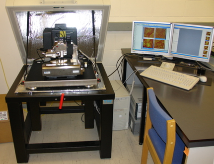
Atomic Force Microscopy: Mapping a Surface with a Tiny Probe
How can a microscope measure the height of features too small to see directly? Atomic force microscopy, or AFM, uses a sharp probe to sense a surface. Its measurement is based on forces and movement, making it a different kind of observation from an ordinary photograph.
The probe and cantilever
Bruker explains that an AFM probe sits at the end of a flexible cantilever. Forces between the tip and specimen cause the cantilever to bend. The instrument detects this deflection and uses it to gather information about the surface.
A common detection arrangement reflects light from the cantilever toward a position-sensitive detector. As the cantilever moves, the detected light position changes. Precise positioning and feedback help the instrument follow the specimen’s topography.
The resulting map connects measured signals with position. It is not produced simply by shining light on the tiny feature and magnifying it. The probe’s interaction with the specimen is central to the method.
Different ways to follow a surface
Bruker’s documentation distinguishes contact mode from tapping approaches. In contact mode, the tip remains in contact while feedback maintains a selected interaction condition. In tapping mode, an oscillating cantilever interacts intermittently with the surface.
The choice matters because the probe can affect the object being measured. Bruker notes that lateral forces in contact mode can distort or displace some specimens. An appropriate imaging mode helps match the measurement to the material.
A practical research example
Imagine a laboratory comparing the surface roughness of two coating formulations. It could scan several representative areas using consistent settings, document the probe and analysis method, and compare the measured height variation. This is a hypothetical study, not a result from a particular coating.
A useful report would also explain the size of the scanned area. One tiny smooth region cannot establish that a whole coated panel is smooth. Repeated measurements and a clear sampling plan make the interpretation more credible.
More than a topographic image
Bruker’s force-spectroscopy guidance describes moving the probe toward and away from the sample while recording interaction. With suitable calibration and analysis, such measurements can investigate properties such as adhesion or stiffness.
That additional capability does not mean every colorful AFM image is a direct map of a single material property. The measurement mode, calibration, probe, and model used for interpretation all matter. Readers should ask what signal was actually recorded and how it was converted into the reported result.
Summary
Atomic force microscopy uses a small probe and flexible cantilever to measure interactions at a surface. It can map topography and support carefully designed force measurements. Its useful precision depends on suitable imaging conditions, calibration, and a clear understanding of the probe’s effect on the specimen.
Works Cited
Bruker. “Atomic Force Microscopy FAQs.” n.d. Accessed 10 Oct. 2026.
Bruker. “Atomic Force Microscopy for Life Sciences.” 2024. Accessed 10 Oct. 2026.
Bruker. “A Practical Guide to AFM Force Spectroscopy and Data Analysis.” n.d. Accessed 10 Oct. 2026.
Zureks. “Atomic force microscope by Zureks.” Wikimedia Commons, 2007. Image. CC BY-SA 3.0. Accessed 10 Oct. 2026.
An atomic force microscope and its control computer. Image: Zureks; CC BY-SA 3.0.