Electron Microscopy: Seeing Structure with an Electron Beam
A material’s visible surface does not reveal all of its important structure. Electron microscopy uses interactions between electrons and a specimen to examine details at much smaller scales. The resulting image can answer useful questions, but it also depends on how the specimen and instrument are prepared.
Two common approaches
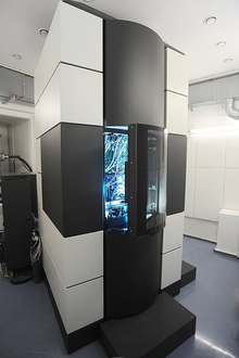
Thermo Fisher Scientific distinguishes scanning electron microscopy, or SEM, from transmission electron microscopy, or TEM. SEM scans a focused beam across a specimen and detects signals produced by its interaction with the surface. TEM uses electrons transmitted through a sufficiently thin specimen to examine internal structure.
The difference is important because the methods answer different kinds of questions. A surface feature and an internal arrangement are not the same object of study. Choosing a microscope begins with deciding what information is required.
How an image is formed
The manufacturer’s SEM explanation describes an electron source, lenses, scanning coils, detectors, and a specimen chamber. The lenses and coils control where the beam goes, while detectors collect signals that can be turned into an image. Different detected signals can emphasize different aspects of the specimen.
The picture is therefore a measured representation, not simply a tiny photograph produced with ordinary light. Imaging settings, detector choice, and specimen properties all influence what is visible. Magnifying the display cannot create detail that the measurement did not resolve.
Preparation can change the answer
Thermo Fisher’s sample-preparation guidance explains that vacuum conditions can affect samples containing moisture or delicate structures. Preparation has to make the specimen suitable for examination while avoiding misleading changes as far as possible.
TEM adds a demanding thickness requirement because electrons must pass through the specimen. A carefully prepared thin section can reveal useful internal information, but the process should not be assumed to leave every material feature untouched.
Imagine a laboratory investigating why a manufactured surface has unexpected defects. It could compare affected and unaffected specimens prepared in the same way, document the imaging conditions, and connect the observed features with other measurements. This is a hypothetical investigation rather than a report of a specific failure.
Benefits and interpretation
Electron microscopy can reveal morphology and structure that other observations miss. However, the examined area may be small compared with the whole component. A striking feature in one image does not establish how common that feature is throughout a production batch.
A useful report includes scale information, preparation details, and the reason the selected images represent the question being studied. Readers should be able to distinguish an observation from a conclusion about its cause.
Summary
Electron microscopy uses electron–specimen interactions to reveal fine detail. SEM and TEM provide different views, and preparation is central to both. The most valuable images are those tied to a clear question, documented conditions, and careful interpretation of what the measurement actually shows.
Works Cited
Thermo Fisher Scientific. “Transmission Electron Microscopy vs Scanning Electron Microscopy.” n.d. Accessed 10 Oct. 2026.
Thermo Fisher Scientific. “What Is Scanning Electron Microscopy?.” n.d. Accessed 10 Oct. 2026.
Luyk, Erik. Thermo Fisher Scientific. “Expert Sample Preparation Techniques for SEMs.” 16 Jan. 2020. Accessed 10 Oct. 2026.
Alice im Miniland. “TITAN.” Wikimedia Commons, 13 May 2011. Image. CC BY-SA 4.0. Accessed 10 Oct. 2026.
A Titan transmission electron microscope. Image: Alice im Miniland; CC BY-SA 4.0.